 Prostate Restored
Prostate Restored
Prostate Restored

 Photo: Tuấn Kiệt Jr.
When the bladder is full, stretch receptors in its muscular wall stimulate a parasympathetic nervous system response. This results in contraction of the detrusor muscle of the bladder and relaxation of the smooth muscle of the internal urethral sphincter. Thus allowing outflow of urine into the urethra.

It's used in ayurvedic medicine as a digestive healing agent. Now Western medicine has begun to study how turmeric can help with gut inflammation...
Read More »
Testosterone levels are typically at their highest in men who are in their late teens and early 20s. That means they respond quickly to sexual...
Read More »
Nine ways to induce urination Tapping the area between navel and pubic bone. ... Bending forward. ... Placing a hand in warm water. ... Running...
Read More »
10 Estrogen Boosting Foods 10 Estrogen building foods: Tofu. Tofu is produced from soy milk which is naturally high in phytoestrogens, specifically...
Read More »Muscles of the pelvic floor and perineum Explore study unit Female perineum Explore study unit Male urinary bladder and urethra Explore study unit Relations The internal urethral sphincter is located at the junction between the urinary bladder and the urethra. Here, supported alongside the bladder neck by the pubovesical ligaments, it surrounds the intramural (preprostatic) part of the urethra and comes in contact with the base of the prostate (males) or the endopelvic fascia (females). Let's recall that the endopelvic fascia is an umbrella term for the connective tissue that envelops the pelvic organs and attaches them to the lateral walls of the pelvis. The external urethral sphincter is contained in the deep perineal space (pouch), which is situated between the pelvic diaphragm and the perineal membrane. It spans the urogenital hiatus of the pelvic diaphragm and fills the areas between the pudendal canals, through which the pudendal nerve and internal pudendal artery and veins pass. The external urethral sphincter is closely related to the levator ani, deep transverse perineal and rectourethral muscles. The puboperineal component of the levator ani muscle, forms an open circle around the external sphincter creating a hiatus at the ventral aspect. The external urethral sphincter is also closely associated with the perineal body, the central tendon of perineum, and may adhere to the body along its posterior aspect. In males, the external sphincter is closely related to the prostate. It sits tightly against the prostate’s apex and is surrounded by a fibrous tissue which is a continuation of the prostate sheath. Semi-circular skeletal muscle fibers at the apex of the prostate blend into those of the external urethral sphincter. The bulbourethral glands are embedded within the sphincter’s muscle fibers. In females, the striated muscle of the external urethral sphincter is closely related to the puborectalis component of the levator ani muscle, while inferiorly it blends with the smooth muscle of the urethra and vagina. [Male urinary bladder] Innervation Pudendal nerve Nervus pudendus Synonyms: Internal pudic nerve, Nervus pudendalis The urethral sphincter complex receives both somatic and autonomic innervation. These supply its voluntary and involuntary components, respectively. The smooth muscle fibers of the internal urethral sphincter receive both sympathetic and parasympathetic innervation. Sympathetic supply arises from the lower thoracic and upper lumbar (T11 - L2) segments of the spinal cord. These nerves maintain tonic contraction of the internal urethral sphincter, thereby preventing urine outflow from the bladder into the urethra. Conversely, the parasympathetic supply arises from sacral levels S2 - S4 (spinal micturition center). It acts to “inhibit” the internal sphincter muscle, thereby relaxing it and allowing urine to pass from the bladder into the urethra. The skeletal muscle fibers of the external urethral sphincter receive somatic motor innervation from the perineal branches of the pudendal nerve (S2-4). These nerves stem from neurons found in an area of the spinal cord called Onuf’s nucleus, which in turn receives fibers from the pontine micturition center of the central nervous system. When stimulated, Onuf’s nucleus acts to increase the resting tone in the external urethral sphincter, the skeletal muscle is contracted and urination is stopped or slowed. Central nervous system inhibition of Onuf’s nucleus results in relaxation of the external urethral sphincter. This facilitates voiding. Blood supply Inferior vesical artery Arteria vesicalis inferior 1/4 Synonyms: Arteria vesicalis caudalis The internal urethral sphincter is supplied by the inferior vesical artery (males) or the vaginal artery (females). The external urethral sphincter receives blood supply from the internal pudendal artery (females) or the artery of bulb of penis (bulbourethral artery), a branch of the internal pudendal artery (males). All of these arteries stem from the anterior division of the internal iliac artery. Function The main action of the urethral sphincter complex is to compress the urethra. This is important as it provides control over urinary continence. The internal sphincter’s smooth muscle resting state is one of contraction or ‘closure’, in which urine is prevented from passing through the internal urethral orifice into the urethra. The external sphincter is also thought to contribute to resting closure, although it’s most important feature is augmenting closure to prevent urination or voluntary opening to allow voiding. During the voiding phase of micturition, both the internal and external sphincters relax to allow urine to pass through the urethra. When the bladder is full, stretch receptors in its muscular wall stimulate a parasympathetic nervous system response. This results in contraction of the detrusor muscle of the bladder and relaxation of the smooth muscle of the internal urethral sphincter. Thus allowing outflow of urine into the urethra. This process is involuntary, however the flow of urine can be voluntarily stopped by contraction of the external urethral sphincter. At an appropriate time and place, the external urethral sphincter can then be voluntarily relaxed, allowing the passing of urine. A secondary function of the urethral sphincter complex is to prevent retrograde ejaculation of semen into the bladder. In males, semen and urine share the urethra as a passageway. Sympathetic stimulation during orgasm in males simultaneously causes contraction of the internal urethral sphincter at the neck of the urinary bladder. This serves to prevent semen from traveling proximally along the urethra, through the internal urethral orifice and into the bladder. In women, the 3 part external urethral sphincter also acts to pull the urethra caudally and inferiorly as well as compress the urethra against the anterior wall of the vagina, aiding urinary continence.

There are several treatment options for an enlarged prostate. You can take alpha-blockers such as terazosin (Hytrin) or tamsulosin (Flomax) to help...
Read More »
Since turmeric (and its derivative curcumin) are not going to show up in your urine, they can not directly affect anything that happens in your...
Read More »
Fluxactive Complete is conveniently packed with over 14 essential prostate powerhouse herbs, vitamins and grade A nutrients which work synergistically to help you support a healthy prostate faster
Learn More »Sources Kenhub does not provide medical advice. You can learn more about our content creation and review standards by reading our All content published on Kenhub is reviewed by medical and anatomy experts. The information we provide is grounded on academic literature and peer-reviewed research.You can learn more about our content creation and review standards by reading our content quality guidelines References: Moore, K. L., Dalley, A. F., & Agur, A. M. R. (2014). Clinically Oriented Anatomy (7th ed.). Philadelphia, PA: Lippincott Williams & Wilkins.

Prostate enlargement tends to come with age. ... Lifestyle Changes Limiting alcohol and caffeine consumption. Reducing liquid consumption before...
Read More »
The “Iron Triangle” in health care refers to the concept that access, cost and quality cannot all be simultaneously improved. The premise is that...
Read More »
Finland. Finland has the highest blond hair population by percentage of the total population. Nearly 80% of the population has blond hair, and an...
Read More »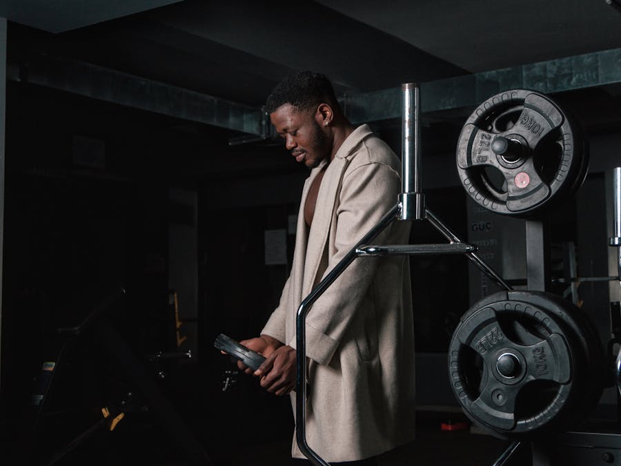
The 4 Phases of Weight Loss Phase -1 – GLYCOGEN DEPLETION. Glycogen Depletion: ... Phase -2 – FAT LOSS. This is the sweet spot for healthy weight...
Read More » Promotion
 Promotion